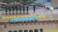
 Modeling mechanics of biological soft structures has been a long time endeavor for functional morphologists as well as theoretical biologists. In general, this field focuses on morphologies without any rigid skeleton (internal or external). They are usually soft tissues supported by some fluid which allows very large deformation. The notion of “hydrostatic skeleton” became well-known by the 50’s largely due to research on worms (cnidarians, annelids, and nematodes). Clark and Cowey established how soft-bodied animals achieved extreme extension with helical reinforcing fibers in the body wall (Clark and Cowey, 1958). The oblique fibers winding around the body allows very large longitudinal stretching. Soon this type of fiber reinforcement was found in many other cylindrical biological structures including those of plants. In 1980’s, a new wave of theoretical investigation of soft-bodied animal locomotion began. Keller and Falkovitz attempted a model of worm crawling using finite difference method which calculated the transverse traveling wave along the body and its associated contact friction (Keller and Falkovitz, 1983). A few years later, Dobrolyubov generalized this line of reasoning to traveling deformation (both transverse and longitudinal). He claimed that the transverse traveling wave can represent caterpillar locomotion while the longitudinal traveling wave resembles crawling worms. Then he gave an example on how this model could describe snake’s locomotion (Dobrolyubov, 1986). These models proposed credible mechanisms for locomotion, but did not explain how animals achieved those body deformations. In another Journal of Theoretical Biology paper, Wadepuhl presented probably the first comprehensive finite element hydrostatic skeleton model based on medical leech which had been well studied by then (Muller et al., 1981; Sawyer, 1986; Stern-Tomlinson et al., 1986; Wadepuhl and Beyn, 1989). This model included geometry, elastic properties of the body wall, internal volume, and body pressure. It revealed some principles of antagonism in worm-like structures as well as the pressure-volume interactions (265 Wadepuhl, M. 1989). At about the same time, Wainwright nicely summarized the mechanics of cylindrical biological structures in his famous little book “Axis and Circumference” (Wainwright, 1988).
Before the turn of the 21st century, Journal of Theoretical biology continued to host models of hydrostatic skeleton. However, experimental data gradually dominated the modeling efforts. Skierczynski et al constructed an updated leech model empirically based on dimensions of animals in limiting cases, passive properties of the tissues, muscle responses to activation, and the transform from motor-neurons to muscles. It assumes elliptical shapes for cross-sections, constant volume, and that the shape tends to minimize the potential energy. It simulates the vermiform elongation and predicts the pressure changes (Skierczynski et al., 1996). Similarly Alscher and Beyn simulated the motion of leech using Lagrangian mechanics and a large system of differential-algebraic equations (Alscher and Beyn, 1998).
While leech models seemed to develop with fast pace, earthworm studies were thriving as well. Dobrolyubov refined his mass transfer wave model and published another paper in JTB with Douchy on peristaltic transport. This general model attempted to explain the digestive transport as well as locomotion by caterpillars, earthworms, snake and snails (Dobrolyubov and Douchy, 2002). Accoto et al added to JTB another earthworm kinematics model, again based on constant volume and simple friction (Accoto et al., 2004). With these numerous hydrostatic skeleton models, it was thought that soft-bodied animal locomotion is more or less realized and what we learned from worms can be applied to others such as caterpillars. Unfortunately, caterpillars are simply not worms in all biomechanical respects.
Caterpillar’s body differs from that of a worm in several essential features: 1) Extension in the longitudinal direction is accounted by numerous inter-segmental folds instead of body wall stretching. 2) Body pressure is highly variable and less predictable. 3) It contains more compressible volume in the body. 4) There is no segmental septum that compartmentalizes the animals. 5) Caterpillars are legged systems with discrete and on-off attachments. As the results, the helical fiber-reinforced cylinder model does not apply. The constant volume assumption does not hold, and real-time pressure recording lacks correlation to body movements. Frictional model based on mass transfer is useless in this system. What’s more, caterpillars don’t move with one single gait and/or body configurations. In this study, we seek an alternative approach to model this worm-like structure that is so much unlike worms.
References
Accoto, D., Castrataro, P. and Dario, P. (2004). Biomechanical Analysis of Oligochaeta Crawling. J. Theor. Biol. 230, 49-55.
Alscher, C. and Beyn, W. J. (1998). Simulating the Motion of the Leech: A Biomechanical Application of DAEs. Numerical Algorithms 19, 1-12.
Clark, R. B. and Cowey, J. B. (1958). Factors Controlling the Change of Shape of Certain Nemertean and Turbellarian Worms. J. Exp. Biol. 35, 731.
Dobrolyubov, A. I. (1986). The Mechanism of Locomotion of some Terrestrial Animals by Travelling Waves of Deformation. J. Theor. Biol. 119, 457-466.
Dobrolyubov, A. I. and Douchy, G. (2002). Peristaltic Transport as the Travelling Deformation Waves. J. Theor. Biol. 219, 55-61.
Keller, J. B. and Falkovitz, M. S. (1983). Crawling of Worms. J. Theor. Biol. 104, 417-442.
Muller, K. J., Nicholls, J. G. and Stent, G. S. (1981). Neurobiology of the Leech: Cold Spring Harbor Laboratory Pr.
Sawyer, R. T. (1986). Leech Biology and Behaviour: Clarendon Press Oxford.
Skierczynski, B. A., Wilson, R. J. A., Kristan Jr, W. B. and Skalak, R. (1996). A Model of the Hydrostatic Skeleton of the Leech. J. Theor. Biol. 181, 329-342.
Stern-Tomlinson, W., Nusbaum, M. P., Perez, L. E. and Kristan, W. B. (1986). A Kinematic Study of Crawling Behavior in the Leech, Hirudo Medicinalis. Journal of Comparative Physiology A: Neuroethology, Sensory, Neural, and Behavioral Physiology 158, 593-603.
Wadepuhl, M. and Beyn, W. J. (1989). Computer Simulation of the Hydrostatic Skeleton. the Physical Equivalent, Mathematics and Application to Worm-Like Forms. J. Theor. Biol. 136, 379-402.
Wainwright, S. A. (1988). Axis and Circumference: The Cylindrical Shape of Plants and Animals. Cambridge: Harvard University Press.


.jpg)