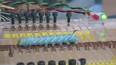
Recently I decided to look at more surface features of Manduca caterpillar bodies with SEM. This time I wanted to explore the folding structures on the soft cuticle.
The first thing I noticed was how hairy these cute caterpillars really are. No wonder people call caterpillars "fussy worms" in tropical Taiwan where I grew up. If vision is weak and proprioception is irrelevant, then tactile sensing must be dominant.
Then I couldn't help focusing my electron beam on the spiracles.... well, they look like tiny stadium to me. This the the hairs around the air slit can't be for tactile functions, or are they?
 I think these are hairs that help repel moisture and particles to keep the air flow smooth. Maybe I should look into the literature.
Finally, I must show you at least one image of the crochets (microscopic claws) on the caterpillar prolegs. They are simply gorgeous!! I got many more images with higher magnification, but it's hard to explain what you are looking at in such close-up photos. This image was actually taken three weeks ago.
 These caterpillars relay on these double array of crochets to grip on to any substrate. When a proleg retracts, these crochets are pulled into the cuticle pocket on the left side of the image. And the whole leg closes like a purse to prevent any unwanted hooking. It's so simple but reliable. I wonder if there is any better strategies for controlling these hooks array with large surface deformation... (another long night of restless dream)

.jpg)
Double array of crochets look like zipperless zipper!
ReplyDelete